
Preparing for MRI-guided Focused Ultrasound, or MRgFUS, is different from preparing for traditional surgery. Because the procedure is incisionless, does not require implanted hardware, and is usually performed without general anesthesia, many patients are surprised by how manageable the process feels. Still, preparation matters. A successful focused ultrasound experience begins well before the treatment day, with careful evaluation, thoughtful planning, and a clear understanding of what the procedure can and cannot do.
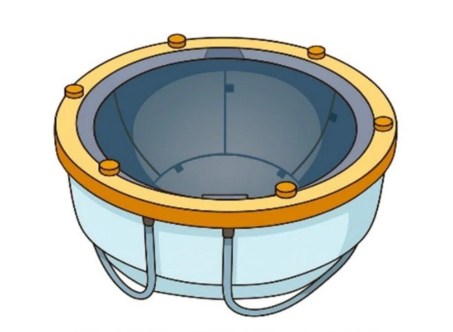
Confirming focused ultrasound is right for you
The first and most important part of preparation happens weeks to months before the procedure: making sure focused ultrasound is the right treatment for you. This begins with confirming the diagnosis and understanding exactly which symptom is causing the greatest difficulty. For many patients, the main issue is disabling tremor from essential tremor or tremor-dominant Parkinson’s disease. In selected patients with Parkinson’s disease, focused ultrasound may also be considered for other motor symptoms depending on the target and the individual clinical situation. The key point is that MRgFUS is not chosen simply because a tremor exists. It is considered when symptoms are significant enough to interfere with daily life—eating, drinking, writing, dressing, working, or maintaining independence—and when medications have not provided enough benefit or have caused side effects.
During this early phase, the movement disorders neurologist plays a central role. Tremor can come from several different conditions, and the right diagnosis is essential because the risks and benefits of MRgFUS depend heavily on the underlying problem. Your neurologist will review your history, examine the tremor carefully, look for signs of Parkinson’s disease, essential tremor, dystonia, ataxia, imbalance, or other neurologic features, and help determine whether focused ultrasound is likely to improve the symptoms that matter most to you. This is also the time to review which medications have been tried, what doses were used, whether they helped, and whether side effects limited treatment.
Imaging and the skull CT
The evaluation also includes imaging. A brain MRI is needed to map your anatomy and help the neurosurgeon plan the target. A specialized CT scan of the skull is also commonly obtained because focused ultrasound beams must pass through the skull to reach the brain target. The CT helps measure skull characteristics, including skull density, to determine whether ultrasound energy can be delivered safely and effectively. This step can feel technical, but in simple terms it tells the team whether the “window” through the skull is favorable for treatment.
Safety steps in the weeks before
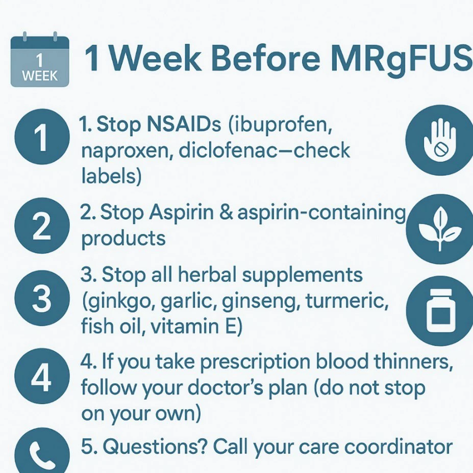
As the procedure gets closer, preparation shifts from candidacy and planning to practical safety steps. In the days to weeks before treatment, your team may review medications, especially blood thinners, antiplatelet drugs, anti-inflammatory medications, or supplements that could increase bleeding risk. Unlike many operations, focused ultrasound usually requires far less preoperative medical testing because there is no incision and no general anesthesia. However, if you have heart disease, lung disease, a history of stroke, or other medical concerns, your team may request additional clearance to make sure the day is as safe as possible.
Preparing emotionally
This is also the time to prepare emotionally. Even though MRgFUS is incisionless, it is still a brain procedure, and it is very common to feel nervous as the date approaches. Many patients worry about the MRI scanner, the head frame, shaving the scalp, whether the procedure will hurt, or whether it will work. These concerns are normal. A good preparation process does not dismiss those fears—it brings them into the open so the team can address them directly. Patients often feel better once they understand the sequence of the day and realize they will be awake, communicating with the team, and actively participating throughout the treatment.
A practical dry run can be very helpful. A few days before the procedure, consider reviewing exactly where you are going, where to park, which entrance to use, and how long it will take to get from the car to the check-in area. If you are traveling from out of town, it may be worth arriving the day before and staying nearby so the morning feels calm rather than rushed. The goal is to remove small frustrations before they become big stressors. On the day of treatment, you want your energy focused on the procedure itself, not on traffic, parking, elevators, or uncertainty about where to go.
What the team is doing behind the scenes
Behind the scenes, the focused ultrasound team is also preparing carefully. Your imaging is reviewed, the target is planned, and the treatment strategy is tailored to your anatomy and symptoms. The MRI, CT scan, treatment software, ultrasound system, and safety checks all come together around one specific patient: you. By the time you arrive, the team has already studied your case and prepared the equipment and plan needed for your treatment.
Arriving on treatment day
On the day of focused ultrasound, you will usually arrive at an MRI suite rather than a traditional operating room. You will change into MRI-safe clothing and remove metal items such as jewelry, watches, hearing aids, phones, or anything that could interfere with the MRI. The team will review the plan, answer last-minute questions, and make sure you are ready before beginning.
The head shave and frame
One of the more emotional parts of the day for many patients is the head shave. This is necessary because the ultrasound waves must pass cleanly through the scalp, and the treatment helmet requires close contact with the head. For some patients, especially those with longer hair, this can feel like a major step. It is important to acknowledge that honestly. Hair grows back, but the moment can still feel vulnerable. Many patients do better when they plan for this in advance with hats, scarves, wigs, or simply time to adjust emotionally.
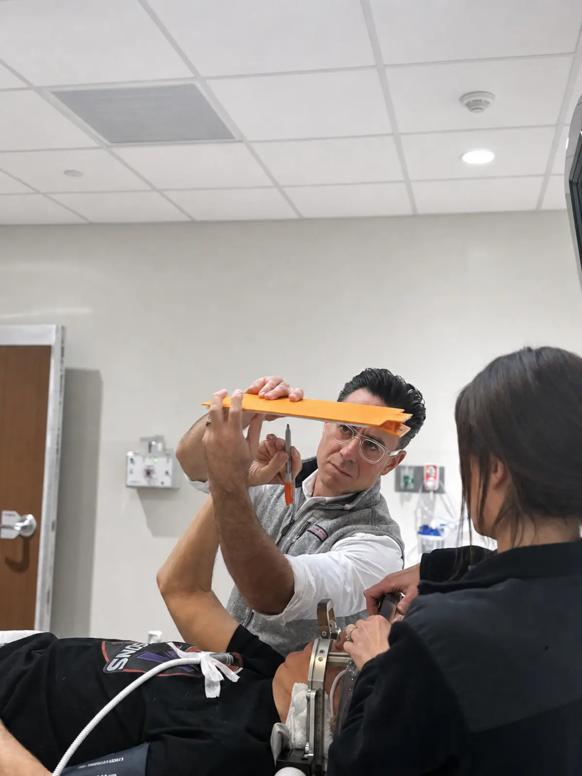
The head frame is then placed to keep your head completely still. Before this is done, the scalp is numbed at four small pin sites. Most patients describe the frame placement as pressure or tightness rather than sharp pain. This is often the part people worry about most beforehand, and many are relieved afterward to find that it was much more manageable than expected.
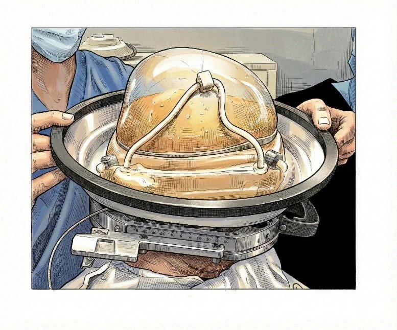
Once the frame is secured, a soft membrane is placed over the scalp, and the head is positioned in the ultrasound helmet. Cool circulating water fills the space between the scalp and the helmet. This water helps transmit the ultrasound beams and protects the scalp from heating. You may feel coolness around the head while the rest of your body is kept warm with blankets. You will then be positioned in the MRI scanner, with monitors in place and a call button in your hand. Even though you are inside the scanner, you are not alone. The team communicates with you throughout the procedure.
The treatment, step by step
The treatment itself is performed step by step. First, the team obtains MRI images and confirms the target. Then low-energy test sonications are delivered. These are essentially practice runs to confirm that the ultrasound energy is reaching the planned spot. After that, the energy is gradually increased in small stages. After each sonication, the team checks your symptoms and asks about side effects. You may be asked to draw spirals, hold a cup, tap your fingers, or perform other simple tasks. This real-time testing is one of the most important advantages of MRgFUS: the team can see whether the tremor is improving while the treatment is happening.
Most patients feel little to no pain during the sonications. Some may feel warmth, pressure, dizziness, nausea, or a brief “hot scalp” sensation, especially if higher energy is needed. These sensations are temporary and are monitored closely. The team can pause, adjust, or provide medication if needed. The treatment continues until the team sees clear symptom improvement and confirms that the target has reached the appropriate therapeutic temperature safely.
Immediately afterward
After the procedure, the head frame is removed, and you are observed in recovery. There are no stitches, no incision, no implanted device, and usually no overnight hospital stay. Most patients go home the same day. For many, the most memorable part of the day is seeing a hand that was shaking become steadier in real time—sometimes while still in the MRI suite.
Recovery
Recovery after focused ultrasound is usually much lighter than recovery after traditional surgery, but it is not nothing. The brain has still been treated, and temporary swelling around the target can cause side effects. The most common issues include imbalance, a tendency to lean, mild headache, scalp soreness at the pin sites, eye or forehead swelling, tingling around the lips or tongue, mild speech changes, fatigue, or temporary clumsiness. These effects often improve over days to weeks, but balance precautions are important early on. Patients should be careful walking, especially at night, on stairs, or on uneven surfaces, and should use a cane, walker, or caregiver support if needed.
Setting expectations
Expectations after MRgFUS should be realistic. The goal is meaningful improvement, not perfection. Tremor often improves substantially, especially in the treated hand, but it may not disappear completely. Focused ultrasound does not cure essential tremor or Parkinson’s disease, and it does not stop the natural progression of the underlying condition. If only one side is treated, the untreated side may still have symptoms. In carefully selected patients, staged second-side treatment may be considered later, but only after the team confirms that speech, balance, and coordination have recovered well from the first side.
Unlike DBS, there is no device to program afterward. That simplicity is one of the major appeals of focused ultrasound. There are no batteries, no wires, no implanted hardware, and no device adjustments. Follow-up remains important, however. Your team will monitor your recovery, help adjust medications if appropriate, and track both benefit and side effects over time.
The larger message is that preparing for focused ultrasound is about more than showing up for a procedure. It is about confirming the right diagnosis, choosing the right target, setting realistic goals, preparing emotionally and practically, and knowing what the recovery will feel like. At Rush, the process is designed so that by the time treatment day arrives, you understand what is happening, why it is being done, and who will be with you at each step.