Deep Brain Stimulation surgery is one of the most precise operations performed in modern neurosurgery. Although patients often think of DBS as “brain surgery,” the procedure itself is best understood as a carefully planned sequence of steps designed to place a very thin electrode into a specific brain network with millimeter-level accuracy. The goal is not simply to place hardware. The goal is to place the DBS lead in the safest and most effective location so that, weeks later, programming can translate that accuracy into meaningful symptom improvement.
At RUSH, DBS surgery begins well before the patient enters the operating room. In the days leading up to surgery, the team reviews the patient’s MRI, confirms the target, studies the safest trajectory, and prepares the navigation, robotic, stereotactic, and imaging systems around that individual patient’s anatomy. This preoperative planning is one of the most important parts of the procedure. The brain is not identical from one person to another, and DBS cannot be approached as a standardized “one-coordinate-fits-all” operation. Modern DBS relies on patient-specific imaging, careful pathway planning, and intraoperative confirmation to reduce risk and maximize accuracy.
The basic goal is the same for both awake and asleep DBS: to place one or two DBS leads into the intended target, such as the subthalamic nucleus, globus pallidus internus, or ventral intermediate nucleus of the thalamus. These targets are small, deep structures involved in motor control. Once the lead is placed, it is connected to an extension wire under the skin, which then connects to a small battery-powered device called an implantable pulse generator, usually positioned under the skin in the upper chest. The system is fully internal. After healing, the neurologist programs the device to deliver stimulation that modulates the abnormal brain network responsible for symptoms such as tremor, stiffness, slowness, dyskinesia, or dystonia.
What Happens Before the Operation Begins
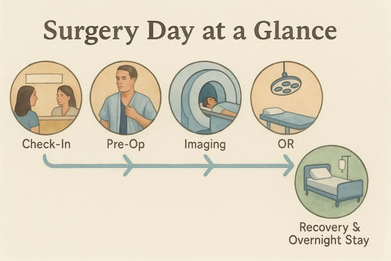
On the morning of surgery, patients arrive in the preoperative area and meet the nursing, anesthesia, and surgical teams. This is often the moment when anxiety is highest. That is normal. Many patients have spent weeks thinking about the operation, and the reality of the day can feel emotional. The team expects this and takes time to review the plan, answer last-minute questions, confirm medications, and make sure both the patient and loved ones understand what will happen next.
The surgery begins with confirmation of the plan. The team verifies the patient’s identity, the surgical target, the side or sides being treated, and the planned approach. Depending on the technique, a stereotactic frame or frameless markers may be placed. If a frame is used, the scalp is numbed first. Patients usually feel pressure rather than sharp pain. In awake DBS, this may be done before going to the operating room. In asleep DBS, some of these positioning and imaging steps may occur after anesthesia has started. For awake frame placement, the scalp is thoroughly numbed and most patients feel pressure, not pain.
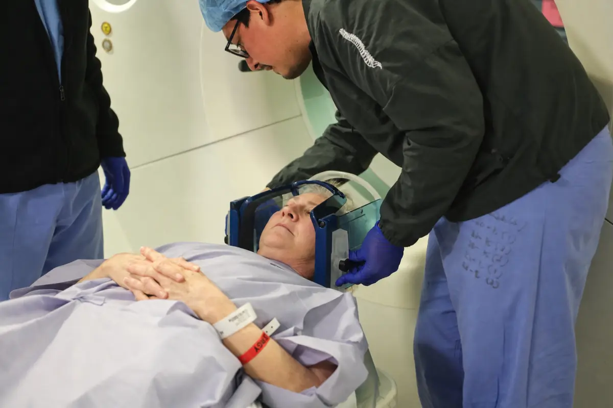
A CT scan may then be obtained and merged with the preoperative MRI. This image fusion creates a three-dimensional coordinate system for the day of surgery. In practical terms, the MRI provides the detailed map of the brain, while the CT confirms the position of the stereotactic system and helps guide the route. The planned path is designed to avoid blood vessels, ventricles, and critical structures while reaching the target as directly and safely as possible.
Once in the operating room, the patient is positioned carefully. The team performs a formal safety “time-out,” confirms the plan again, and prepares the scalp with sterile antiseptic solution. Only a small amount of hair is trimmed when possible. The surgical field is then draped. Before the incision is made, local anesthetic is injected so the scalp remains comfortable. Even in asleep DBS, local anesthetic is often used to reduce postoperative discomfort.
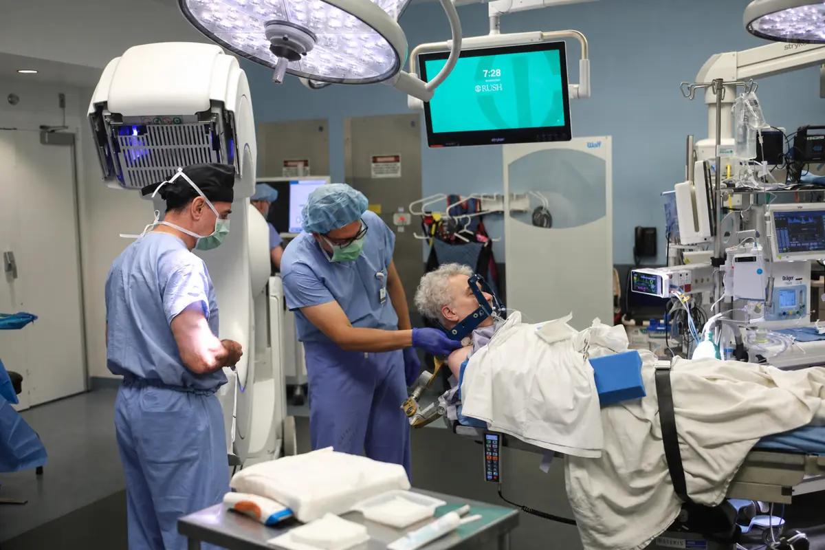
The opening in the skull is small—often about the size of a nickel. Through this opening, the surgeon advances the instruments along the planned trajectory. This is where the two main approaches begin to differ.
Two Roads to the Same Destination: Awake and Asleep DBS
DBS can be performed using an awake approach or an asleep approach. Both are accepted, modern techniques. Both can produce excellent outcomes in experienced hands. The difference is not the goal; the difference is how the team confirms the target and how the patient experiences the operation.
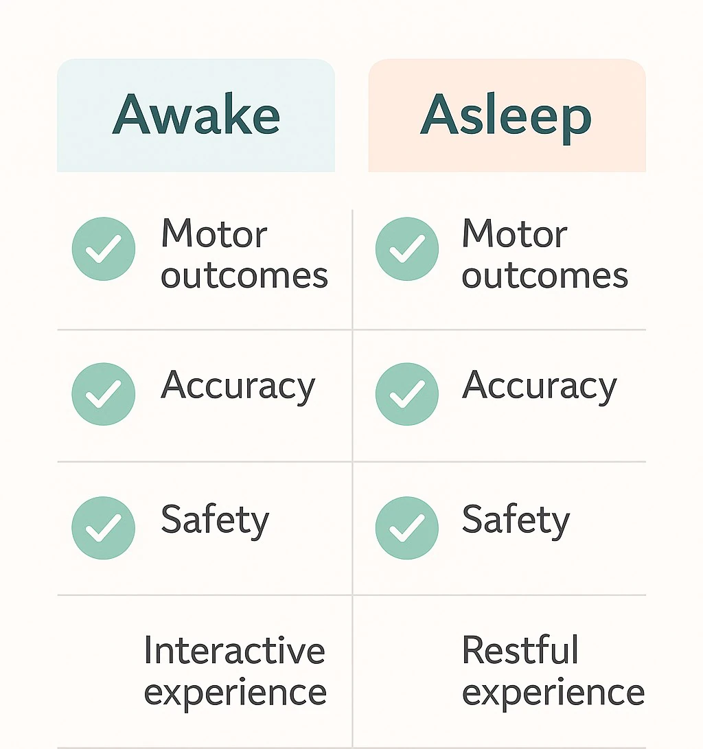
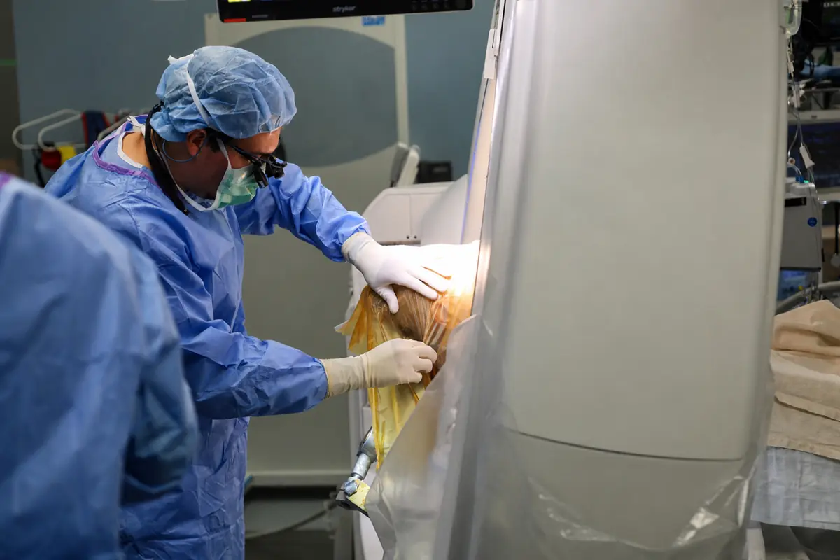
Awake DBS uses physiologic confirmation. The patient is awake for key parts of the surgery, allowing the team to use microelectrode recording and test stimulation. Microelectrode recording, often called MER, allows the team to “listen” to the activity of individual brain regions as the electrode approaches the target. Different targets have characteristic firing patterns. Test stimulation then allows the team to observe benefit and side effects in real time. Awake DBS is described as using MER to fine-tune the target and brief stimulation testing while patients perform simple tasks such as opening and closing the hand or speaking a phrase.
Asleep DBS uses image-based confirmation. The patient is under general anesthesia, and the surgeon relies on high-resolution MRI, computer-guided navigation, stereotactic or robotic guidance, and intraoperative imaging to confirm lead placement. Advances in MRI visualization and intraoperative CT or MRI have made asleep DBS increasingly common. A review from RUSH notes that modern neuroimaging has supported a shift toward direct, image-based targeting under general anesthesia, with asleep DBS showing reliable targeting accuracy and similar short-term clinical efficacy compared with awake approaches in published retrospective comparisons.
The literature does not support a simplistic claim that one approach is universally better. A 2024 systematic review and meta-analysis including 19 studies and 1,900 patients evaluated clinical outcomes, surgical outcomes, complications, quality of life, mood, and cognitive measures in awake versus asleep DBS for Parkinson’s disease. A 2020 robot-assisted DBS study found that asleep and awake approaches both improved motor outcomes, with asleep surgery showing shorter procedure duration and similar electrode accuracy and short-term improvement. The most accurate way to present this to patients is that both approaches are valid, and the best choice depends on the patient, target, anatomy, symptoms, anxiety level, and the expertise of the center.
Awake DBS: Mapping the Target With the Patient’s Participation
Awake DBS has a long history and remains an excellent approach, especially when the surgical team wants physiologic confirmation of the target or when real-time symptom testing is particularly valuable. The word “awake” can sound intimidating, but patients are not awake in the way many imagine. The scalp is numbed, sedation is used strategically, and the team talks the patient through each step. Many patients doze during portions of the case and are awakened only when participation is needed.
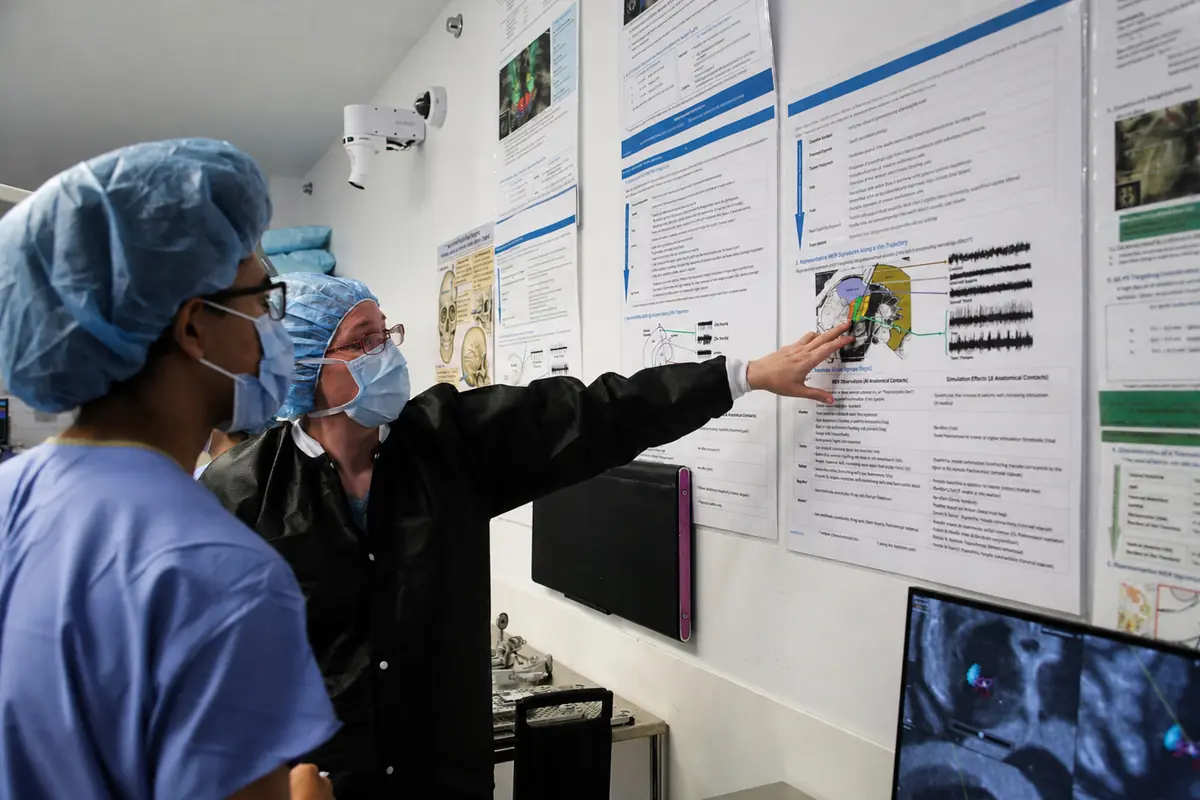
After the patient is positioned and the small skull opening is made, the surgeon advances a very fine recording electrode toward the target. This is the microelectrode recording phase. The recording electrode is much thinner than the permanent DBS lead and allows the team to identify the electrical signature of the brain structures along the path. Different targets—such as STN, GPi, or VIM—have different patterns of activity. This is described as “listening” to the brain to guide the last millimeters, with patients sometimes hearing soft pops or crackles through a speaker.
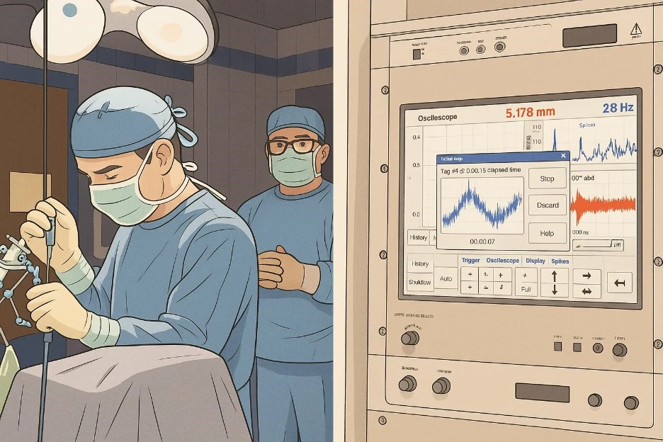
Once the team identifies the best physiologic location, test stimulation is performed. This is often the most memorable part of awake DBS. A tremor may quiet. A rigid hand may loosen. A patient may open and close the hand more easily. At the same time, the team intentionally checks for side effects, such as tingling, pulling in the face or hand, speech changes, or imbalance. These effects are temporary and stop when stimulation is reduced. This testing helps define the “therapeutic window,” meaning the range where stimulation provides benefit without unacceptable side effects.
After testing confirms the best location, the temporary recording electrode is removed and the permanent DBS lead is placed. The lead is secured, imaging may be obtained to confirm its position, and the scalp is closed. If both sides are being treated, the process is repeated on the other side.
In many awake DBS workflows, the brain leads are placed during the first procedure, while the extension wires and pulse generator are placed later in a second surgery, usually about a week later. This second stage is performed asleep. Through a small incision behind the ear, the lead is connected to an extension wire that travels under the skin down the neck to the chest, where it connects to the battery. The behind-the-ear connector and chest generator are positioned in a deeper, well-padded plane to sit as flush and discreetly as possible.
The rationale for awake DBS is straightforward: it allows physiologic confirmation and real-time testing. This can be reassuring for both patient and surgeon, especially when anatomy is complex or when symptom testing is important. Some patients also find it powerful to witness their tremor improve during the operation. For others, the thought of being awake is stressful, and that is where asleep DBS may be a better fit.
Asleep DBS: Precision Through Imaging, Navigation, and Intraoperative Confirmation
Asleep DBS is performed under general anesthesia. The patient goes to sleep at the beginning of the operation and wakes up when the procedure is complete. For patients who are anxious about being awake, have severe symptoms when medications are held, have difficulty lying still, or prefer a single-stage operation, asleep DBS can be a very attractive option.
The procedure begins with anesthesia and careful positioning. Once the patient is asleep, the surgical team secures the head, obtains or confirms imaging, and uses the preoperative MRI and intraoperative CT or MRI guidance to navigate to the target. The scalp is prepared, a small incision is made, and a small skull opening is created. The DBS lead is then advanced along the planned trajectory to the target.
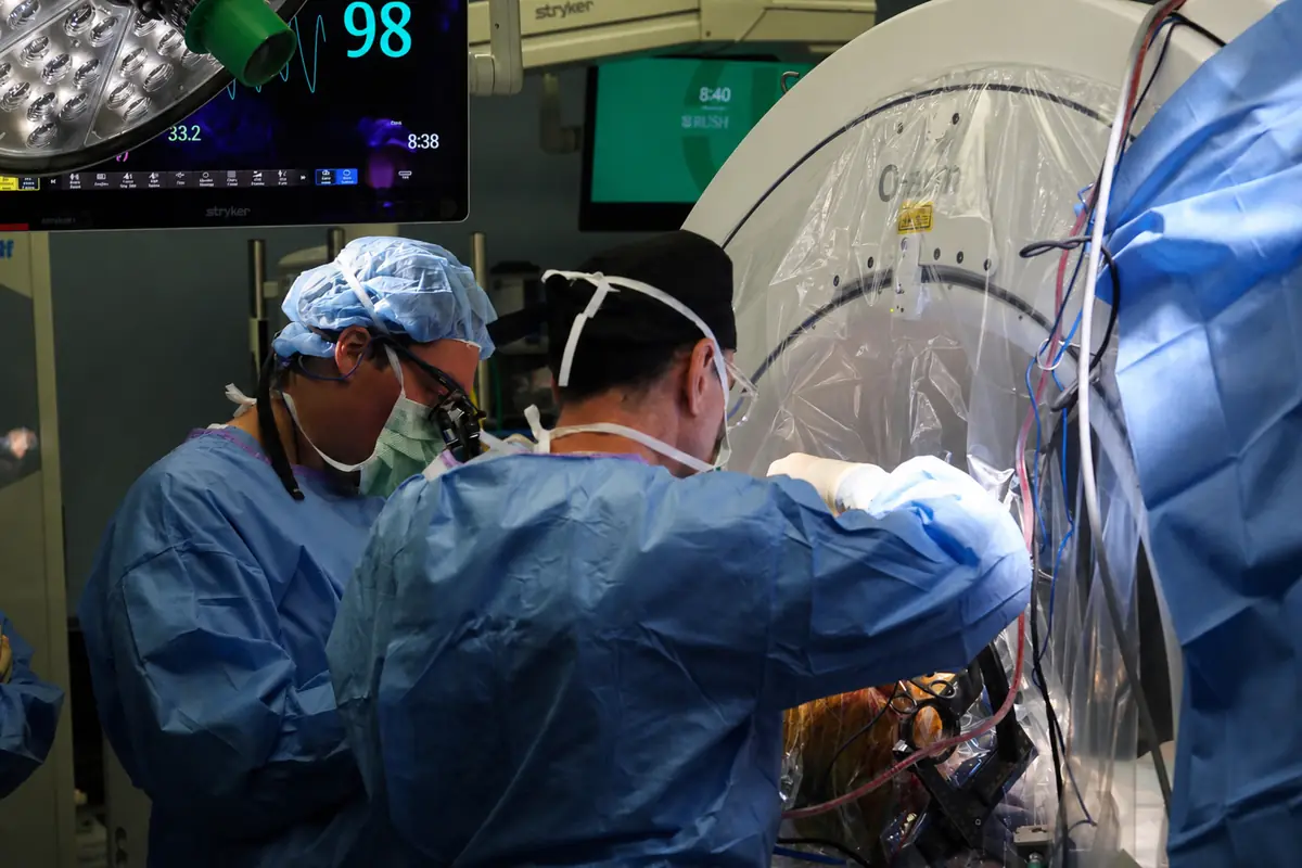
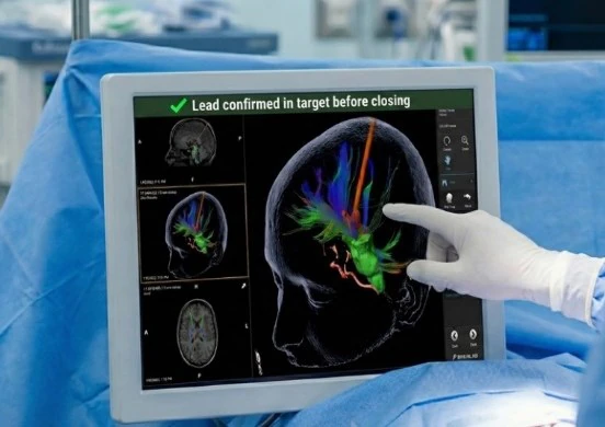
The difference is that the team does not rely on the patient performing tasks during the procedure. Instead, accuracy is confirmed with imaging. Intraoperative CT or MRI allows the team to see the position of the lead relative to the planned target before the operation is finished. A RUSH-authored technical review describes asleep DBS as using direct image-based targeting, often with intraoperative CT or intraoperative MRI, and notes that these methods have demonstrated reliable targeting accuracy for STN and GPi DBS.
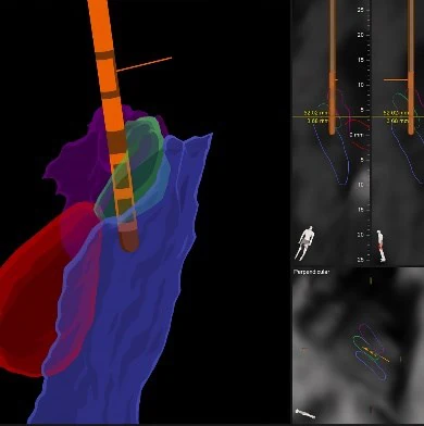
Asleep DBS is described as using advanced imaging and navigation to place the lead and then obtaining an intraoperative scan with the lead in place, allowing the team to reconstruct the images in three dimensions and confirm the tip and contact positions relative to the planned target. If a small adjustment is needed, it can be made before the procedure is completed.
In many asleep DBS cases, all hardware can be placed in one session: the brain leads, extension wires, and chest battery. This avoids a separate second-stage procedure and can simplify the logistics for patients and families. After the leads are confirmed, the extension wires are tunneled beneath the skin from the scalp, behind the ear, and down the neck to the chest. The pulse generator is placed under the skin below the collarbone, and the system is tested electrically before closure.
Cosmetic and comfort considerations matter. The connector behind the ear is positioned to reduce prominence when possible, and the chest battery is placed in a location that considers comfort, body habitus, clothing, seatbelt position, and prior scars or devices. Patients should understand that the battery may still create a small visible or palpable contour, especially in thinner individuals, but the goal is to make it low-profile, comfortable, and safe.
The rationale for asleep DBS is also straightforward: it allows the entire procedure to be done under anesthesia, often in one stage, using modern imaging and navigation to confirm accuracy. It can reduce the emotional burden of awake surgery and may be better suited to patients who cannot tolerate medication withdrawal, prolonged positioning, severe anxiety, or active symptoms during surgery. Published comparisons suggest that asleep DBS can achieve similar short-term motor outcomes to awake DBS when performed by experienced teams using modern imaging.
How We Decide Between Awake and Asleep DBS
The decision between awake and asleep DBS is individualized. It is not a matter of bravery, and it is not a test of toughness. Some patients are excellent candidates for awake DBS and appreciate the reassurance of intraoperative testing. Others are better served by asleep DBS because it is more comfortable, logistically simpler, or medically more appropriate.
The target also matters. For Parkinson’s disease, both STN and GPi can often be visualized with modern MRI techniques, making image-guided asleep DBS a strong option in appropriate patients. For essential tremor, the VIM target is more challenging because it is not always directly visible on standard MRI, so some centers still prefer awake testing for VIM DBS, although imaging and tractography-based approaches continue to evolve. The RUSH technical review notes that STN and GPi are commonly visualized with modern MRI sequences, while VIM targeting historically relied more on atlas-based coordinates, with tractography of tremor pathways offering promise.
Patient comfort matters as well. Claustrophobia, severe OFF symptoms, painful dystonia, anxiety, inability to lie flat, sleep apnea considerations, and medication needs can all influence the choice. Both approaches are excellent and that the team recommends the one that best fits the patient’s symptoms, anatomy, and comfort.
At a high-volume center, the most important factor is not simply awake versus asleep. It is whether the team can select the right approach for the right patient and execute it with precision. Modern DBS requires expertise in imaging, anatomy, physiology, device technology, programming, and long-term movement disorders care.
Immediately After Surgery
After surgery, patients wake up in the recovery area. A nurse monitors blood pressure, comfort, neurologic status, and the incisions. Patients may have a mild headache, scalp tenderness, or soreness near the chest battery if it was placed that day. Most pain is modest and controlled without opioids.
Most patients spend one night in the hospital. This is primarily for observation and reassurance. The care team checks neurologic function, restarts medications unless instructed otherwise, encourages gentle movement, and monitors for early issues. Most patients go home the next morning.
It is common to feel tired for several days. Some swelling around the scalp or forehead can occur. A subtle bump behind the ear from the connector may be felt under the skin. These are expected parts of healing. The first month is about healing, clean wounds, gentle movement, and realistic pacing, with programming beginning around four weeks later.
When the Benefit Begins
The DBS system is usually not turned on immediately. At RUSH, initial programming typically begins about four weeks after surgery. This waiting period allows swelling to settle and the brain to recover before stimulation is adjusted. During this time, patients generally remain on their usual medications.
Some patients experience a temporary improvement before programming begins. This is known as the microlesion or honeymoon effect. It happens because the placement of the lead itself temporarily disrupts the abnormal circuit. It can be encouraging, but it is not the true long-term DBS effect and it does not happen in everyone.
The true benefit begins with programming. The neurologist activates the device, tests settings, watches for benefit and side effects, and gradually builds a program tailored to the patient. For Parkinson’s disease and essential tremor, improvement may be seen early in programming. For dystonia, benefit often unfolds more slowly over months.
The Central Message
DBS surgery is precise, deliberate, and highly individualized. Whether performed awake or asleep, the operation is designed around the same principles: careful planning, safe trajectory selection, accurate lead placement, confirmation of the target, thoughtful hardware positioning, and long-term programming by an expert movement disorders team.
For patients, the procedure can feel intimidating because it involves the brain. But in experienced hands, DBS is a carefully choreographed operation with a small physical footprint and a large potential impact. The surgery creates the foundation. Programming turns that foundation into therapy. Long-term care sustains the benefit.