
MRI-guided Focused Ultrasound is one of the most technologically advanced procedures in modern neurosurgery. Unlike traditional surgery, there are no incisions, no implanted devices, and no opening of the skull. Instead, the procedure uses highly focused sound waves combined with real-time MRI guidance to target and treat abnormal brain circuits with remarkable precision. Although the physical footprint of the treatment is much smaller than conventional surgery, the planning, technology, and coordination behind it are extraordinarily sophisticated.
For patients, one of the most surprising aspects of focused ultrasound is that they remain awake throughout the procedure. This is not because the treatment is painful, but because remaining awake allows the team to evaluate symptoms in real time and carefully monitor neurologic function as treatment progresses. The procedure is highly interactive, deliberate, and gradual. Every step is designed around safety, precision, and confirming that the treatment is helping the symptom being targeted.
Before the Procedure Begins
The focused ultrasound process begins well before the patient enters the MRI suite. In the days leading up to treatment, the team reviews MRI and CT imaging in detail, analyzes skull characteristics, confirms the treatment target, and prepares the treatment plan specifically for that patient’s anatomy.
One of the unique aspects of focused ultrasound is that the skull itself becomes part of the procedure. Unlike DBS, where instruments physically enter the brain, focused ultrasound must deliver energy through the intact skull. Because skull thickness and density vary between individuals, specialized CT analysis is performed beforehand to evaluate how efficiently ultrasound energy will travel through the skull. This information helps determine both candidacy and procedural planning.
On the day of treatment, patients arrive at the hospital several hours before the procedure begins. The morning is structured and methodical. After check-in, the treatment team reviews the plan again and answers any remaining questions. Although patients are understandably nervous at this stage, many describe feeling calmer once the process becomes organized and stepwise.
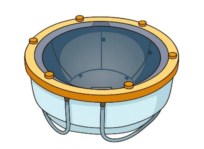
One of the first major steps is shaving the scalp completely. This is necessary because the ultrasound helmet must make direct contact with the scalp to allow the sound waves to pass efficiently and safely through the skull. A flexible membrane and circulating cooled water system are then used during treatment to help conduct the ultrasound energy and regulate scalp temperature.
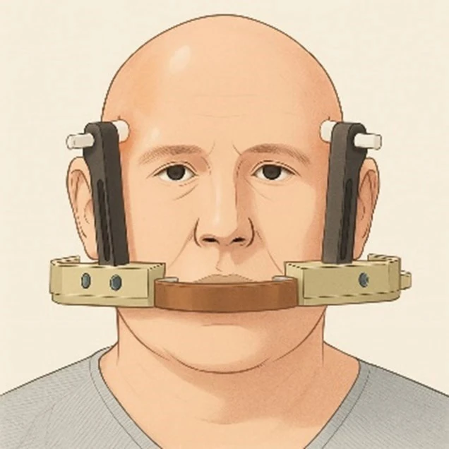
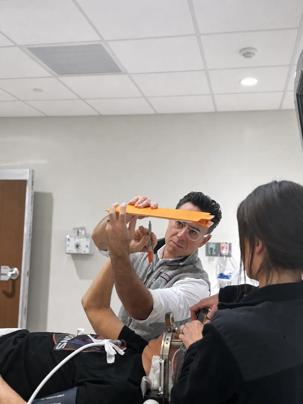
An IV is placed, and the scalp is cleaned carefully. After numbing medication is injected into specific areas of the scalp, a lightweight stereotactic frame is secured to the head. The frame is critically important because it keeps the head perfectly still and allows the treatment to be delivered with submillimeter precision. Although the concept of the frame can sound intimidating beforehand, most patients tolerate it very well and describe it more as a sensation of pressure rather than pain.
Entering the MRI Suite
Once preparation is complete, the patient is brought into the MRI suite and positioned on the MRI table. The focused ultrasound helmet is connected to the head frame, and the patient’s head is surrounded by a water-filled treatment dome containing more than one thousand individual ultrasound transducers.
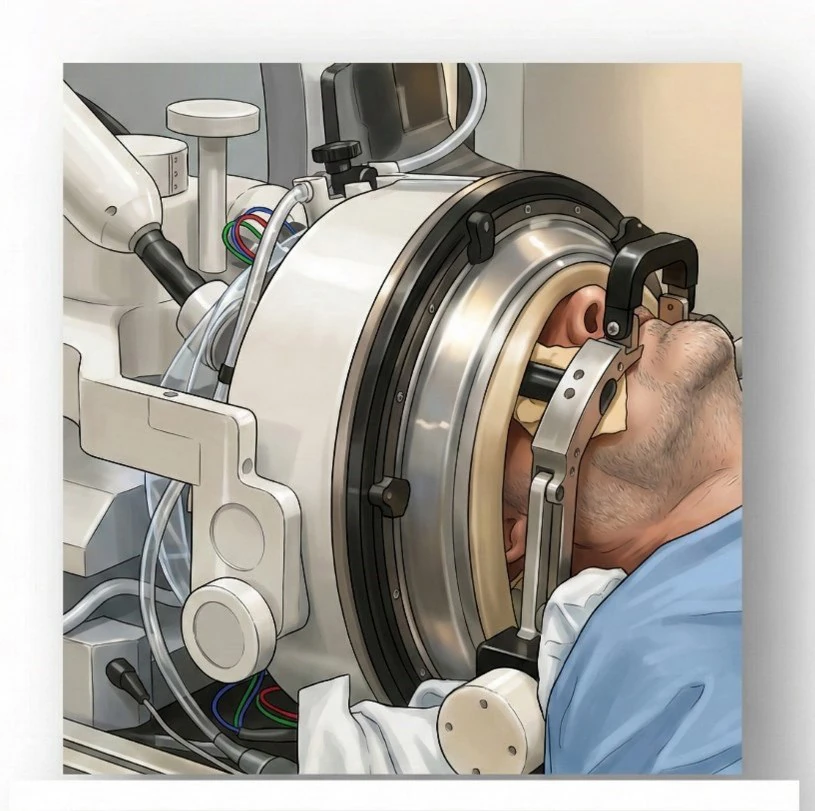
At this stage, the treatment environment resembles a combination of advanced imaging, neurosurgery, and engineering. The MRI scanner provides continuous anatomical visualization, while the focused ultrasound system calculates and delivers energy with extraordinary precision.
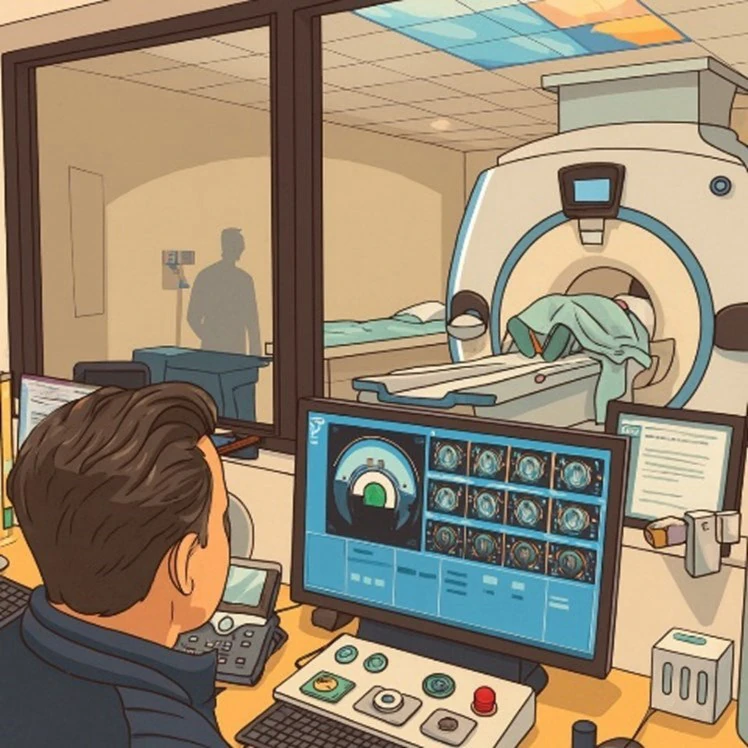
Patients remain awake and communicate continuously with the team throughout the procedure. This ongoing interaction is one of the defining features of focused ultrasound and one of the reasons the treatment can be adjusted so carefully in real time.
Targeting the Brain
Before therapeutic treatment begins, the team first performs detailed MRI imaging to confirm the exact anatomy and treatment target. For essential tremor and tremor-dominant Parkinson’s disease, this most commonly involves the ventral intermediate nucleus (VIM) of the thalamus, a key relay station within the tremor network. Other Parkinson’s-related targets may include structures such as the globus pallidus internus (GPi) or pallidothalamic pathways depending on the indication being treated.
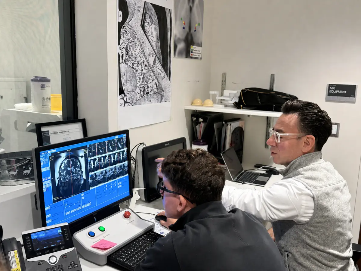
The treatment plan is individualized to each patient’s anatomy. Millimeter-level adjustments matter enormously in functional neurosurgery. The goal is to target the abnormal motor circuit precisely while avoiding nearby structures involved in speech, sensation, balance, or vision.
Once the target is confirmed, the procedure begins with low-energy test sonications. These early sonications do not create permanent tissue effects. Instead, they allow the team to confirm that the ultrasound energy is reaching the intended location safely and accurately.
MRI thermometry—a remarkable feature of the system—allows the team to monitor temperature changes within the brain in real time. This means the surgeon can actually see where heat is developing during treatment and confirm whether adjustments are needed.
The Stepwise Treatment Process
Focused ultrasound is not performed all at once. It is a progressive, carefully staged process.
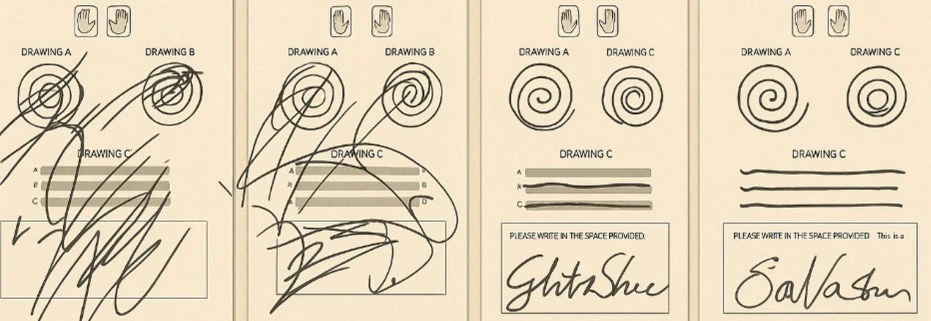
The procedure begins with low temperatures and gradually advances in a series of sonications, each delivering slightly more energy than the last. After each sonication, the team stops and evaluates the patient carefully. Patients may be asked to:
-
draw spirals,
-
hold a cup,
-
touch their finger to their nose,
-
write their name,
-
speak,
-
move their limbs,
-
or perform other simple neurologic tasks.
This real-time testing is one of the most powerful aspects of focused ultrasound. The team is not simply relying on imaging—they are actively observing whether tremor improves and whether any side effects appear.
For many patients, this becomes the most emotional part of the procedure. Tremor that has been present for years—or decades—may suddenly quiet during testing. Patients often see their handwriting improve immediately or notice they can hold a cup steadily for the first time in years. These moments can be profound, both for patients and families.
As temperatures gradually increase into the therapeutic range, a small controlled lesion is created at the target. The lesion itself is tiny, but because it interrupts a critical abnormal circuit, the functional effect can be dramatic.
Throughout treatment, safety remains the overriding priority. If side effects such as numbness, speech changes, imbalance, or unusual sensations appear during lower-energy testing, the target can be adjusted before permanent treatment is delivered.
What Patients Feel During Treatment
Most patients tolerate focused ultrasound very well. The procedure itself is not typically painful, though patients may feel:
-
pressure from the head frame,
-
warmth during sonications,
-
mild scalp discomfort,
-
occasional dizziness,
-
or a brief sensation of imbalance or tingling.
The MRI environment can feel long or mentally tiring, especially since the procedure often lasts several hours from preparation through completion. However, patients are continuously monitored and supported by the team throughout the process.
The treatment is very controlled and deliberate. Nothing happens suddenly or unexpectedly. The patient remains in constant communication with the team, and treatment can be paused whenever necessary.
Completing the Procedure
Once satisfactory tremor control is achieved and the final treatment is completed, the patient is removed from the MRI scanner and the stereotactic frame is taken off. Patients are then monitored for a period of observation.
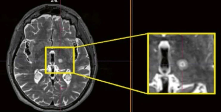
At this point, many patients immediately notice the difference in tremor. Some experience mild fatigue, temporary imbalance, or a sensation of being “off” as the brain begins adapting to the treatment. Mild headache or scalp soreness may also occur from the frame placement.
Unlike DBS, there is no implanted hardware, no chest battery, and no programming phase afterward. The treatment effect comes directly from the lesion itself.
Most patients go home the same day.
The Immediate Recovery Period
The first several days after focused ultrasound are usually straightforward, though patients are encouraged to take things slowly initially. Fatigue is common. Temporary imbalance or gait unsteadiness may occur, particularly during the first days to weeks, as mild swelling develops around the treatment area.
Some patients experience temporary numbness or tingling involving the lips, tongue, or fingers. Others may notice subtle changes in balance or coordination. These effects are usually transient and improve gradually over time as swelling resolves.
One of the major advantages of focused ultrasound is the relative simplicity of postoperative care. There are no wounds to manage, no hardware maintenance, and no programming visits. Most follow-up can be performed through telemedicine, which is particularly helpful for patients traveling from outside the Chicago area.
The Bigger Picture
Focused ultrasound represents a major shift in how neurosurgical movement disorder treatment can be delivered. It combines advanced imaging, engineering, and neuromodulation principles into an incisionless procedure capable of producing immediate functional improvement.
At the same time, the technology itself is only one part of the process. Successful focused ultrasound depends heavily on:
-
accurate diagnosis,
-
careful patient selection,
-
detailed imaging analysis,
-
precise targeting,
-
and extensive experience with movement disorders and neurosurgical anatomy.
At RUSH, focused ultrasound is approached with the same philosophy that guides all advanced neuromodulation care: thoughtful evaluation, individualized treatment planning, technical precision, and long-term patient-centered follow-up. The goal is not simply to perform the procedure, but to achieve meaningful and lasting improvement safely and predictably for the right patient.