
MRI-Guided Focused Ultrasound
Tremor treatment without an incision
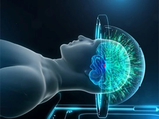
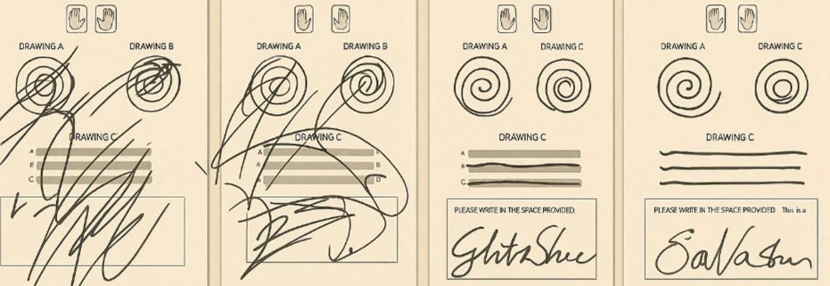
More than a thousand low-energy sound beams pass through the skull and converge on a single point deep in the brain. Patients stay awake throughout, drawing spirals between sonications, and many see their tremor settle before they leave the scanner.
How it works
Sound, focused to a few millimeters
A single ultrasound beam passing through the skull is far too weak to have any effect. Send a thousand of them from different angles and each travels harmlessly through bone and healthy tissue — but where they intersect, the energy is sufficient to raise the temperature of a target a few millimeters across.
Two developments made this practical. MRI shows the target in detail, and MRI thermometry measures temperature at that target in real time, so energy can be raised gradually and stopped the moment the intended effect is reached.
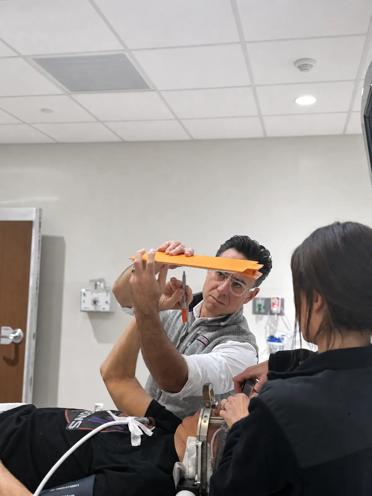
Because patients remain awake, the team tests between sonications — drawing a spiral, holding a cup, touching the nose, speaking a sentence. Benefit and side effects are both measured before anything permanent is done.
What focused ultrasound treats
Approved indications, and expanding
Focused ultrasound was the first incisionless brain procedure for a movement disorder approved in the United States, and the list of approved targets has continued to grow.
FDA approved 2016
Essential Tremor
The most established indication. Thalamotomy of the ventral intermediate (VIM) nucleus for tremor no longer controlled by medication, with staged treatment of the second side now possible in selected patients.
FDA approved 2018, expanded since
Parkinson's Disease
Approved first for tremor-dominant Parkinson's disease, then for other motor symptoms, then for pallidotomy in 2025 and staged bilateral pallidothalamic tractotomy later that year.
Assessed individually
Complex tremor syndromes
Some tremors fit neither essential tremor nor Parkinson's disease. The question is not simply whether tremor is present, but whether reducing it would improve function without worsening speech, coordination or balance.
Treatment day
What happens, step by step
-
1
Before the day
Evaluation and a CT of the skull
The skull forms part of the treatment system, so a CT is used to calculate the skull density ratio — a measure of how efficiently sound will pass through the bone. Alongside it: confirmation of diagnosis, a medication review and high-resolution MRI. The evaluation process →
-
2
Morning of treatment
Head shaved, frame placed
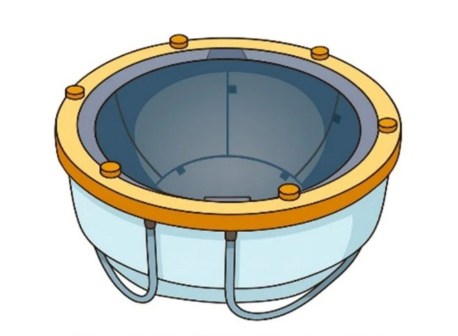
The scalp is shaved completely so nothing scatters the beams. Pin sites are numbed with local anesthetic and a frame holds the head still. An elastic membrane is placed over the scalp and attached to the transducer once you are positioned on the MRI table.
-
3
In the scanner
Test sonications, then treatment
The team begins well below therapeutic energy, confirms the spot is correct, then works upward in steps. Between each one you draw, write, speak and move, so benefit and side effects are measured directly rather than assumed. The procedure in detail →
-
4
Same day
Frame off, and home
There is no incision to heal, no hardware, no battery and no programming appointments. Mild imbalance, tingling or fatigue in the first days is common and usually settles. Much of the follow-up can be done closer to home. Recovery →
Focused ultrasound is permanent
This is the central trade-off and it is worth understanding before deciding. Focused ultrasound creates a small permanent lesion, which means no hardware and no maintenance — but also that a persistent side effect cannot be programmed away as a DBS side effect usually can. Read the outcomes and risks in full, or compare focused ultrasound with DBS.
Go deeper
Everything about focused ultrasound
- What is focused ultrasound
- Who is a candidate
- Conditions treated
- Evaluation
- Preparing for treatment
- Traveling to RUSH Oak Brook
- The procedure
- Recovery
- Outcomes and risks
- Bilateral treatment
- MRgFUS vs DBS
- Life after treatment
- Why RUSH
- Patient education
- Focused ultrasound FAQs
- Trusted external resources
Many patients begin with a video visit
If you live outside Chicago, the first step usually does not require travel. A telemedicine consultation can establish whether focused ultrasound is worth pursuing before you plan a trip.